Keratoacanthoma
What do you do when a bump like this suddenly shows up on your skin and grows fast? It’s not a spot, not a wart, and not quite a skin cancer either. It might be something called a keratoacanthoma — and today we’re going to unpack what that really means.
What Is a Keratoacanthoma?
A keratoacanthoma (KA) is a skin lesion that:
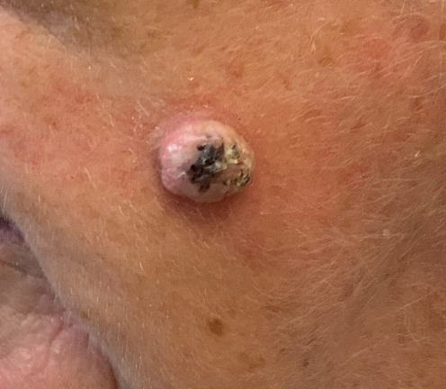
Grows quickly, often over a few weeks, from a small papule into a dome‑shaped lump that has a central crater or keratin plug. PCDS describes that rapid evolution over about 12 weeks. Primary Care Dermatology Society
Has a “volcano‑like” appearance: smooth shoulders, fairly symmetrical, with a central crust or plug of keratin. Primary Care Dermatology Society+2DermNet®+2
Is often firm to the touch, maybe red or skin‑coloured. Because of its features, it lies in a kind of grey zone: it looks like cancer in some ways, but many will eventually shrink and even disappear. DermNetNZ says that although many KAs regress, clinically they may be indistinguishable from well‑differentiated squamous cell carcinoma (SCC). DermNet®
Typical locations are sun‑exposed skin: face, arms, hands, scalp. And almost always in older people (over 60) with fair skin.
Causes & Risk Factors
From what leading dermatology sources tell us, risk factors include:
Sun / UV exposure (chronic).
Age – older age increases risk; most patients are over 60.
Sex – more common in men than women.
Immunosuppression (e.g. transplant recipients, immunosuppressive therapy).
Skin injury, trauma or scars – KAs can appear in sites of previous injury or scarring.
Rare genetic syndromes or variants.
Some drug treatments have also been implicated (e.g. BRAF inhibitors used in melanoma) and very occasionally exposure to PUVA etc.
Why It’s Tricky: Red Flags & Look‑Alikes
Keratoacanthomas are often confused with squamous cell carcinoma (SCC). The similarities are many: rapid growth, crusting, location on sun‑exposed skin. Because of this overlap, many clinicians treat suspected KAs similarly to SCC until proven otherwise. DermNet®+2Patient+2
Here are some red flags or features that may suggest that something more serious than a classic KA is happening:
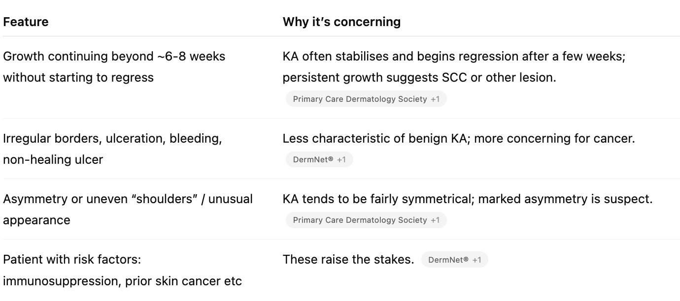
LDiagnosis usually requires a biopsy or excision to check the cells under microscope, because clinically there can be no definitive way to distinguish KA vs SCC.
Outcome & Natural Course
Many KAs regress spontaneously over time (typically over 4‑6 months), leaving a scar. DermNetNZ suggests solitary KAs follow that kind of course. DermNet®
That said, the course is unpredictable. Some KAs do not regress, some may behave aggressively, and because of the risk of misdiagnosis (i.e. a hidden SCC), waiting can carry risk.
Prognosis after excision is excellent; recurrence is uncommon if completely removed.
Treatment: What to Do & When
Because of the diagnostic uncertainty (KA vs SCC), the default is often to treat rather than observe unless you're very confident. Sources like PCDS, DermNet, BAD agree. Primary Care Dermatology Society+2DermNet®+2
Here are the options:
Surgical excision with margins: the gold standard. Removes the lesion, allows histological examination, ensures margins are clear.
Curettage + cautery or curettage + electrodessication: scraping away the lesion then destroying the base. Useful if excision is difficult.
Cryotherapy (freezing) – in small lesions or those in more accessible / less risky sites. British Association of Dermatologists+1
Topical therapies (e.g. 5‑fluorouracil, imiquimod) or intralesional injections (e.g. methotrexate, bleomycin, steroids) in selected situations: e.g. patient medically unfit for surgery, multiple lesions, or cosmetically challenging location.
For very rare, widespread forms (like generalized eruptive KA), systemic treatments (retinoids etc) may be considered. DermNet®+1
Watch‑and‑wait (“wait for spontaneous regression”) is possible, but risky unless the diagnosis is certain or lesion very small and in a non‑cosmetic/non‑risk area. In practice many clinicians choose to remove early to avoid the chance of missing an SCC. DermNet®+2British Association of Dermatologists+2
Summary / What You Should Know
If you see a fast‑growing, volcano‑shaped bump (dome with a crater / central plug), especially on sun‑exposed skin, in someone older, think of keratoacanthoma — but also think of SCC.
The appearance can be very similar to cancer; you can’t reliably tell them apart just by sight. Biopsy or surgical excision is often needed.
Most KAs are treatable, many regress naturally, but the risk of misdiagnosis means medical review is wise.
Early removal tends to give excellent outcomes with minimal recurrence.
What You Should Do If You Suspect One
See a healthcare professional (dermatologist or GP with dermatology interest) especially if the lesion is growing fast, bleeding, changing or won’t heal.
Don’t delay more than a few weeks if red flags present. Many UK guidelines (e.g. PCDS) suggest urgent referral under the 2‑week skin cancer “red flag” pathway for suspected KA/SCC. Primary Care Dermatology Society
Protect your skin: sun protection, avoid burns, be alert to new lumps.
Share this post on:



Copyright © 2024 Dr Finbars' Skin Clinic All rights reserved.